
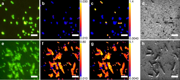
 Fig. 1: 13C and 15N incorporation in representative microbial cells. figure1 Cells from incubations of U1365 9H-3 with 13C-bicarbonate and 15N ammonium (a–d) and 13C,15N-Amino acid mix (e–h). (a, e) SYBR Green I-stained cells under fluorescence microscopy. b, c, f, g Ratio images of 13C/12C (b, f) and 12C15N/12C14N ratios (c, g) of the same regions imaged in a, e, demonstrating locations of 13C and 15N incorporation. Color-scale ranges of the ratios are shown as numbers appearing at top and bottom of the color bar. The background membrane region, which is identified by fluorescence images, is excluded from the ratio calculation and shown as black background. d, h. Secondary electron (NanoSIMS) images of the same regions in a, e. Bars represent 5 µm. Similar images were processed for obtaining the dataset (Supplementary Data 1) of substrate incorporations for 6986 individual cells.